A groundbreaking international collaboration has fundamentally challenged the long-held scientific understanding of how the brain processes visual information, revealing a previously unknown neuron type that drastically enhances our ability to perceive complex objects. For decades, neuroscience textbooks have taught that the initial stages of visual processing in the cortex relied primarily on two types of cells specialized in detecting "edges"—sharp transitions between light and dark. However, an international team, spearheaded by researchers from Stanford University and the University of Göttingen, leveraging advanced artificial intelligence (AI) techniques, has shattered this established model with the discovery of a third, distinct class of neuron in the mouse visual cortex. This newly identified neuron possesses a sophisticated, two-part receptive field, capable of simultaneously recognizing both fine textures and specific spatial arrangements, thereby providing a far more efficient mechanism for distinguishing complex objects from their backgrounds than simple edge detection alone could offer. The findings, published in the esteemed journal Nature Neuroscience, mark a significant paradigm shift in our comprehension of visual perception and hold profound implications for the future of AI and neurological research.
The Foundation of Visual Science: From Edges to Complexity
For over half a century, the field of visual neuroscience has largely been built upon the seminal work of David Hubel and Torsten Wiesel, whose pioneering research in the 1950s and 60s earned them a Nobel Prize. Their investigations into the cat and monkey visual cortices identified two primary types of neurons: "simple cells" and "complex cells." Simple cells were found to respond selectively to edges or bars of light at a specific orientation and location within their receptive field—the particular area of the visual field to which a neuron responds. Complex cells, in contrast, also responded to edges of a specific orientation, but were less sensitive to their exact position, exhibiting a degree of spatial invariance. This "edge detection" paradigm became the cornerstone of visual processing theory, suggesting that the brain constructs an understanding of the visual world by first identifying these fundamental contours and then progressively building up more complex representations.
This model, while immensely influential and explanatory for many basic visual phenomena, has faced inherent limitations when attempting to account for the brain’s remarkable ability to effortlessly segment complex objects from cluttered backgrounds. Imagine a bird perched in a leafy tree, or a face partially obscured by shadows. The brain doesn’t just see a collection of edges; it instantly recognizes the bird or the face as a coherent entity, distinct from its surroundings. This robust object recognition in dynamic and noisy environments suggested that the visual cortex might employ more sophisticated mechanisms than a purely hierarchical processing of simple luminance changes. The challenge, however, lay in uncovering these elusive neural mechanisms, often hidden amidst the intricate network of millions of neurons in the visual cortex. Traditional experimental methods, while powerful for investigating known cell types, were not ideally suited for systematically discovering entirely novel computational strategies employed by neurons.
Unveiling the Third Neuron Type: A New Mechanism for Object Recognition
The breakthrough came through the innovative application of machine learning, specifically deep neural networks, to analyze the activity of mouse neurons. The international team discovered a third class of neuron whose functional properties diverge significantly from the classic simple and complex cells. These newly identified neurons possess a unique "bipartite receptive field," meaning their response area is divided into two distinct components, each specializing in different aspects of visual information.
One part of this receptive field is exquisitely tuned to detect textures. This involves processing "high spatial frequencies," which correspond to dense patterns, fine details, and sharp lines—think of the intricate patterns of fur, feathers, bark on a tree, or the weave of fabric. This component allows the neuron to identify the characteristic surface properties of an object or background. The other part of the receptive field, however, responds to specific spatial arrangements of patterns, often characterized by "low spatial frequencies." Low spatial frequencies represent coarse patterns, larger uniform areas, and the overall structure of an object—such as the distinctive arrangement of a nose and mouth on a face, or the broad outline of an animal.
The crucial insight is how these two parts work in concert. While simple and complex cells are primarily activated by differences in brightness (luminance edges), these new neurons respond to more abstract "edges" defined by differences in texture or spatial frequency. This dual specialization provides a powerful mechanism for functional bipartite invariance, allowing the brain to efficiently separate an object from its background. For instance, if an animal is camouflaged against a similarly colored background, its texture might differ subtly, or its overall shape might create a distinct low-frequency pattern against the background’s high-frequency texture. These new neurons are perfectly poised to detect such nuanced distinctions, enabling rapid and robust object segmentation even in challenging visual scenes. As Professor Andreas Tolias from Stanford University summarized, "Classic simple and complex cells are tuned to simple edges defined by differences in brightness. In contrast, the two-part neurons we found respond to more complex information about edges — that is, differences in texture or spatial frequency. These are precisely the kinds of signals needed to separate an object from its background."
The AI Revolution in Neuroscience: Digital Twins and Discovery
This discovery would have been exceedingly difficult, if not impossible, without the advent of sophisticated artificial intelligence and machine learning techniques. The researchers employed deep neural networks, similar to those used in advanced AI models for image recognition, to create "digital twins" of individual mouse neurons. These digital twins are computational models that accurately predict the activity of real neurons. The process involved feeding vast amounts of visual data into these AI models, allowing them to learn the intricate response characteristics of each neuron.
The University of Göttingen played a pivotal role in developing these digital twins. Professor Fabian Sinz, from Göttingen University’s Institute of Computer Science, highlighted the necessity of this approach: "Neural networks are essential tools for discovering new properties from large data sets — such as these novel neuronal properties." The power of these digital twins lies in their ability to be probed systematically with an almost infinite array of synthetic visual stimuli. Researchers could present millions of different images to the digital twin in seconds, identifying precisely which patterns and characteristics maximally activated a given neuron. This iterative process of generating "varied exciting inputs" (VEIs) allowed them to hypothesize about the specific features these neurons were encoding, far beyond what could be practically tested in vivo with traditional methods.
A critical aspect of the research was the rigorous experimental verification of the AI’s predictions. Professor Alexander Ecker, also from Göttingen, emphasized this point: "The predicted best images are not fantasies of our AI model." He further stressed, "Targeted experiments in real mouse brains, led by researchers at Stanford University, have confirmed the properties predicted by our model are real." This rigorous validation loop—from large-scale in vivo recordings to predictive AI models, then to in silico experiments with AI, and finally back to in vivo verification—represents a powerful new paradigm in neuroscience. It demonstrates how AI can serve as a potent discovery engine, guiding experimentalists to uncover previously hidden biological truths. This interdisciplinary approach, combining advanced computational methods with precise neurophysiological experiments, exemplifies the cutting edge of modern neuroscience.
A Paradigm Shift Confirmed: Researcher Perspectives
The findings represent more than just an incremental addition to our knowledge of the visual system; they signify a genuine paradigm shift. For decades, the field was largely comfortable with the "edge detector" analogy for the early visual cortex. This new discovery forces a re-evaluation of that foundational understanding, suggesting that even at the earliest stages of visual processing, the brain is performing more complex computations than previously imagined. It implies that the visual system isn’t merely identifying lines and contrasts but is actively engaged in sophisticated feature extraction tailored for object segmentation.
The "Key Questions Answered" section from the original source provides valuable insights into how the researchers themselves view the significance: "We knew the basics! For 50 years, we thought the visual cortex was just an ‘edge detector.’ But this study shows it’s much more like a high-tech photo editor. It doesn’t just see lines; it sees the difference between the ‘texture’ of a sweater and the ‘shape’ of the person wearing it simultaneously." This vivid analogy underscores the advanced nature of the newly discovered neuronal function. It highlights a system that is not passively receiving light information but actively parsing and interpreting it in a highly organized and efficient manner from the very first cortical stages.
The implications for the broader neuroscience community are substantial. This research opens up entirely new avenues for investigation. Scientists will now look to explore the prevalence of these neurons across different species, including humans, and to understand their connectivity within the broader visual hierarchy. How do these texture- and arrangement-sensitive neurons communicate with other parts of the brain responsible for higher-level object recognition, memory, and decision-making? The discovery necessitates a re-examination of existing models of visual processing and prompts new theories about the computational strategies employed by the brain to construct our rich visual experience.
Beyond the Lab: Broadening the Impact
The ramifications of this discovery extend far beyond the confines of basic neuroscience research, promising significant advancements in several applied fields.
Advancing Artificial Intelligence
One of the most immediate and profound impacts will be on the field of artificial intelligence, particularly in computer vision. Current computer vision systems, despite their impressive capabilities, still often struggle with robust object recognition in highly cluttered or visually ambiguous environments—challenges that biological vision systems handle with ease. Many contemporary AI models are still fundamentally inspired by the older "edge detection" paradigm, even if implemented with vastly more complex neural networks.
By understanding and mimicking the mechanisms of these newly discovered two-part neurons, AI developers could design more biologically plausible and, crucially, more efficient and robust computer vision algorithms. Imagine autonomous vehicles that can more reliably distinguish pedestrians from complex backgrounds, medical imaging systems that better segment tumors from healthy tissue, or robotics that can identify and manipulate objects in dynamic, unpredictable settings. The ability to simultaneously process texture and spatial arrangement, and to use these differences to define object boundaries, is a critical step towards building AI systems that perceive the world with a sophistication closer to that of biological brains. As noted in the FAQ, "Most computer vision today is still based on the old ‘edge detection’ model. By mimicking these newly discovered two-part neurons, we could build AI that is much better at identifying objects in messy, cluttered environments—just like a mouse (or a human) does." This research provides a direct blueprint for enhancing AI’s perceptual capabilities.
Insights into Neurological Health and Disease
The deeper understanding of fundamental visual processing also carries significant implications for neurological health and disease. Many neurological and psychiatric conditions, such as autism spectrum disorders, schizophrenia, and certain types of dyslexia, involve atypical visual processing. If these newly identified neurons play a crucial role in efficient object segmentation, dysfunctions in their activity or connectivity could contribute to the visual perceptual difficulties experienced by individuals with these conditions.
This discovery could pave the way for new diagnostic tools, allowing clinicians to identify specific deficits in texture- and arrangement-based visual processing. Furthermore, it could inform the development of novel therapeutic interventions, targeting these specific neural circuits to improve visual perception. For individuals with visual impairments, a more detailed understanding of how the brain constructs visual information could lead to the design of more effective visual prosthetics or brain-computer interfaces, which could leverage the brain’s natural processing strategies more directly.
The Future of Visual Neuroscience
This research also highlights the transformative power of interdisciplinary collaboration, particularly at the intersection of neuroscience and computational science. The "inception loop paradigm," as described in the abstract—iterating between large-scale recordings, predictive models, in silico experiments, and in vivo verification—is a powerful methodology that will undoubtedly accelerate discoveries across many domains of neuroscience.
Future research will undoubtedly focus on several key areas. Scientists will seek to determine if similar bipartite receptive fields exist in other parts of the visual system or in other sensory modalities. Comparative studies across different species will be crucial to understand the evolutionary conservation and specialization of these neurons. Furthermore, dissecting the precise molecular and cellular mechanisms underlying the formation and function of these neurons will be a vital next step. Ultimately, by unraveling the intricate computational strategies of the brain, we move closer to a comprehensive understanding of consciousness itself, and how the brain crafts our subjective reality from the raw data of the senses.
Conclusion
The discovery of a third, functionally distinct neuron type in the visual cortex marks a pivotal moment in neuroscience. By challenging a decades-old dogma and revealing a more sophisticated mechanism for object-background separation, this international team has not only deepened our understanding of the brain’s incredible efficiency but also opened new frontiers for artificial intelligence and therapeutic interventions. The successful integration of cutting-edge AI with rigorous neurophysiological experimentation demonstrates a powerful new approach to scientific discovery, promising an era of unprecedented insights into the mysteries of the brain. The brain is not merely an "edge detector"; it is a marvel of parallel processing, capable of extracting complex, multi-dimensional information from the visual world, allowing us to navigate and understand our surroundings with remarkable ease.
Research Details:
Author: Melissa Sollich
Source: University of Göttingen
Contact: Melissa Sollich – University of Göttingen
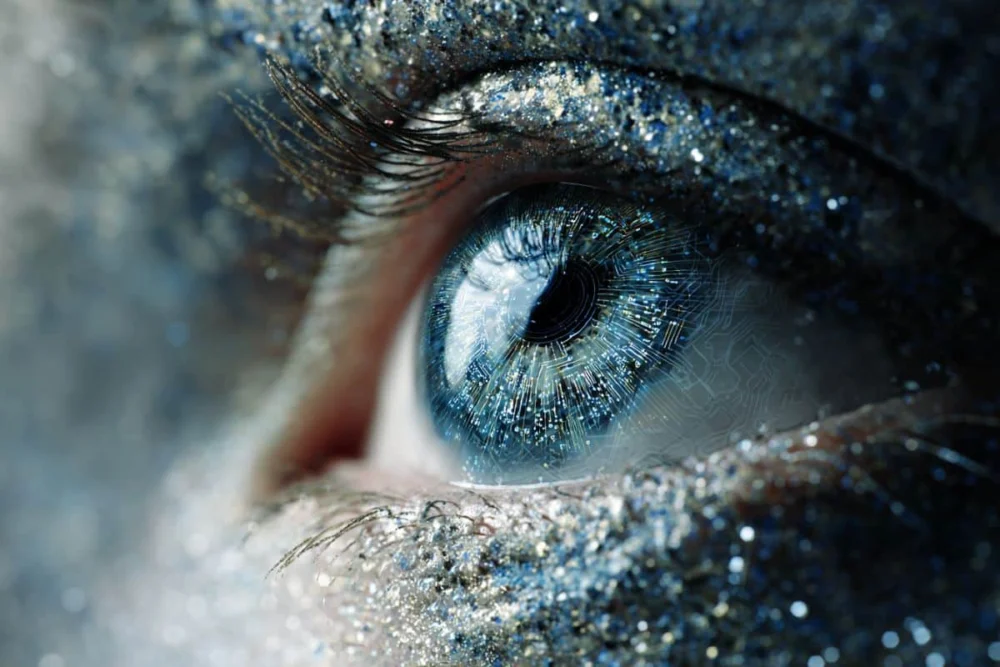
Image: The image is credited to Neuroscience News
Original Research: Open access.
“Functional bipartite invariance in mouse primary visual cortex receptive fields” by Zhiwei Ding, Dat Tran, Kayla Ponder, Zhuokun Ding, Rachel Froebe, Lydia Ntanavara, Paul G. Fahey, Erick Cobos, Luca Baroni, Maria Diamantaki, Eric Y. Wang, Andersen Chang, Stelios Papadopoulos, Jiakun Fu, Taliah Muhammad, Christos Papadopoulos, Santiago A. Cadena, Alexandros Evangelou, Konstantin Willeke, Fabio Anselmi, Sophia Sanborn, Jan Antolik, Emmanouil Froudarakis, Saumil Patel, Edgar Y. Walker, Jacob Reimer, Fabian H. Sinz, Alexander S. Ecker, Katrin Franke, Xaq Pitkow & Andreas S. Tolias. Nature Neuroscience
DOI:10.1038/s41593-026-02213-3
Abstract
Functional bipartite invariance in mouse primary visual cortex receptive fields
Sensory systems support generalization by representing features that persist under input variation; however, identifying the neuronal basis of these invariances remains difficult due to high-dimensional and nonlinear neural computations.
Here we leverage the inception loop paradigm, iterating between large-scale recordings, predictive models and in silico experiments with in vivo verification, to characterize neuronal invariances in mouse primary visual cortex (V1). We synthesize varied exciting inputs (VEIs), dissimilar images that drive target neurons.
These VEIs revealed a new bipartite invariance: one subfield encodes a shift-tolerant high-frequency texture and the other encodes a fixed low-frequency pattern. This division aligns with object boundaries defined by spatial frequency differences in highly activating images, suggesting a contribution to segmentation.
Analysis of the MICrONS dataset revealed a hierarchy of excitatory neurons in mouse V1 layers 2/3: postsynaptic neurons exhibited greater invariance than their presynaptic inputs, while neurons with lower invariance formed more connections.
Together, these results provide insights and scalable methodology for mapping neuronal invariances.