A groundbreaking multi-center international study has unveiled a critical vulnerability in modern healthcare: neither highly experienced radiologists nor advanced multimodal large language models (LLMs) can consistently differentiate between artificial intelligence (AI)-generated "deepfake" X-rays and genuine medical images. This unsettling discovery, published today in Radiology, a journal of the Radiological Society of North America (RSNA), exposes a significant threat with far-reaching implications, from the potential for widespread medical fraud and litigation to sophisticated cybersecurity attacks capable of destabilizing digital health records and compromising patient care.
The research, involving 17 radiologists across 12 institutions in six countries—the United States, France, Germany, Turkey, the United Kingdom, and the United Arab Emirates—systematically evaluated their ability to identify synthetic X-rays created by leading AI models, including OpenAI’s ChatGPT (specifically GPT-4o) and Stanford Medicine’s open-source generative AI diffusion model, RoentGen. Even when explicitly forewarned about the presence of synthetic images within the datasets, radiologists achieved an average accuracy of only 75% in distinguishing real from fake. This modest success rate underscores a profound challenge for the medical imaging community and the broader healthcare ecosystem, signaling an urgent need for advanced protective measures and specialized training.
Deepfake X-Rays: A Growing Threat to Diagnostic Integrity
The study’s findings highlight a burgeoning concern: the increasing sophistication of generative AI models means that digitally fabricated medical images are becoming virtually indistinguishable from authentic ones to the human eye, even that of a trained specialist. This blurring of reality and simulation poses an unprecedented risk to the integrity of medical diagnoses, patient safety, and the foundational trust placed in medical documentation.
Dr. Mickael Tordjman, lead study author and a post-doctoral fellow at the Icahn School of Medicine at Mount Sinai, New York, articulated the gravity of the situation. "Our study demonstrates that these deepfake X-rays are realistic enough to deceive radiologists, the most highly trained medical image specialists, even when they were aware that AI-generated images were present," he stated. He further emphasized the "high-stakes vulnerability for fraudulent litigation if, for example, a fabricated fracture could be indistinguishable from a real one." Beyond civil disputes, Dr. Tordjman pointed to the "significant cybersecurity risk if hackers were to gain access to a hospital’s network and inject synthetic images to manipulate patient diagnoses or cause widespread clinical chaos by undermining the fundamental reliability of the digital medical record."
The Study’s Methodology and Unsettling Results
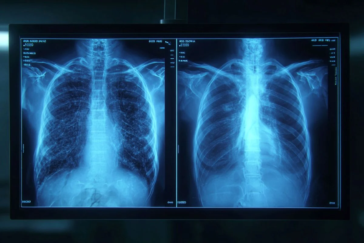
The research employed a rigorous methodology, presenting radiologists with 264 X-ray images, half of which were authentic clinical images and half generated by AI. The study was structured into distinct phases and datasets to thoroughly assess detection capabilities.
In the initial phase, radiologists were presented with a dataset containing a mix of real and ChatGPT-generated images spanning multiple anatomical regions. Crucially, they were blinded to the study’s true purpose and asked to assess image quality and provide diagnoses. During this phase, only 41% of the participants spontaneously identified any images as being AI-generated, even when prompted if they noticed anything unusual after evaluating the technical quality of the ChatGPT images. This low initial detection rate underscores the insidious nature of these deepfakes when observers are not specifically looking for them.
Upon being informed that synthetic images were indeed present in the dataset, the radiologists’ mean accuracy in differentiating real from synthetic X-rays improved to 75%. Individual performance varied considerably, ranging from a low of 58% to a high of 92%. This variability suggests that while some radiologists possess a more refined ability to spot these fakes, a significant portion remain highly susceptible to deception.
A second dataset focused exclusively on chest X-ray images, with half being authentic and the other half generated by RoentGen, a model specifically designed for medical image synthesis. In this scenario, radiologist accuracy in detecting the synthetic images ranged from 62% to 78%.
Human vs. AI: A Battle for Authenticity
The study also pitted advanced multimodal LLMs against the deepfake images, mirroring the human radiologist challenge. Four prominent LLMs were tested: GPT-4o (OpenAI), GPT-5 (OpenAI), Gemini 2.5 Pro (Google), and Llama 4 Maverick (Meta). Their performance in identifying AI-generated X-rays varied, with accuracy rates ranging from 57% to 85% for the ChatGPT-generated images. Interestingly, even ChatGPT-4o, the very model used to create some of the deepfakes, was unable to detect all of its own creations, though it outperformed Google and Meta LLMs in identifying them. For the RoentGen synthetic chest X-rays, the LLM models’ performance ranged from 52% to 89%. This indicates that even cutting-edge AI, while capable of generating these convincing fakes, struggles to reliably distinguish them from authentic images, suggesting a complex challenge that transcends current technological capabilities.
The research also delved into the demographic factors influencing detection rates. Surprisingly, there was no statistically significant correlation between a radiologist’s years of professional experience and their accuracy in detecting synthetic X-ray images. This finding suggests that seasoned veterans are just as vulnerable to deepfake deception as their less experienced counterparts. However, musculoskeletal radiologists demonstrated significantly higher accuracy than other radiology subspecialists, perhaps due to their specific expertise in identifying subtle structural abnormalities in bone and joint imaging, areas where AI-generated images often exhibit tell-tale "perfection."
The Alarming Stakes: Fraud, Misdiagnosis, and Cybersecurity Chaos
The implications of these findings are profound and multi-faceted, threatening to undermine the very pillars of medical practice and patient trust. The "deepfake" phenomenon, broadly defined as any video, photo, image, or audio recording that appears real but has been created or manipulated using AI, has moved from entertainment and political disinformation into the high-stakes realm of healthcare.
Vulnerability in Litigation and Insurance
One of the most immediate and tangible risks lies in fraudulent litigation and insurance claims. Imagine a scenario where a fabricated X-ray depicting a severe fracture or an undiagnosed pathology is introduced as evidence in a personal injury lawsuit or an insurance claim. If even expert radiologists cannot definitively discern its artificial nature, the legal system could be compromised, leading to unjust settlements, increased healthcare costs, and a breakdown of forensic integrity. The ability to "hallucinate" a perfectly convincing injury on an X-ray could become a powerful tool for bad actors seeking illicit financial gain, making it exceedingly difficult for legal and insurance professionals to verify the authenticity of medical evidence.
A New Vector for Cyberattacks in Healthcare
Beyond fraud, the study highlights a critical cybersecurity vulnerability. Healthcare institutions are increasingly reliant on digital medical records (DMRs) and picture archiving and communication systems (PACS). A sophisticated cyberattack could involve not just data exfiltration or ransomware, but the insidious injection of synthetic images into patient records. Such an attack could create "widespread clinical chaos," as Dr. Tordjman warned. Fabricated images could lead to misdiagnoses, unnecessary surgeries, delayed essential treatments, or even the withholding of care based on a falsified "clear" X-ray when a patient genuinely suffers from a serious condition like pneumonia or cancer. The potential for patient harm, coupled with the erosion of trust in digital medical records, represents an existential threat to modern healthcare.
Anatomy of a Deepfake: Identifying the "Too Perfect" Image
Despite their deceptive realism, deepfake X-rays often possess subtle, characteristic imperfections that, upon close scrutiny, can betray their artificial origin. Dr. Tordjman explained, "Deepfake medical images often look too perfect." He elaborated on these tell-tale signs: "Bones are overly smooth, spines unnaturally straight, lungs overly symmetrical, blood vessel patterns excessively uniform, and fractures appear unusually clean and consistent, often limited to one side of the bone."
These "perfect" anomalies stem from the generative AI models’ learning process. They are trained on vast datasets of real images and attempt to synthesize new ones by identifying and replicating common patterns. In doing so, they can inadvertently smooth out the natural variations, asymmetries, and subtle irregularities inherent in biological structures. A genuine X-ray, for instance, might show slight spinal curvatures, asymmetrical lung vascularity, or nuanced bone textures that are challenging for current AI to perfectly replicate without introducing an unnatural uniformity. While these features are difficult for human radiologists and even other AI models to consistently identify, they represent the critical clues that future detection tools must be designed to recognize.
The Broader Context: Generative AI’s Double-Edged Sword in Medicine
The emergence of deepfake X-rays is part of a larger narrative surrounding the rapid advancement of generative artificial intelligence. In recent years, models like ChatGPT, DALL-E, and Midjourney have captured global attention with their astonishing ability to create realistic text, images, and even videos from simple prompts. These technologies are built upon complex neural networks, often employing architectures like Generative Adversarial Networks (GANs) or diffusion models, which learn to mimic real-world data distributions.
The Ascent of Generative Artificial Intelligence
The journey of generative AI from theoretical concept to widespread application has been swift. Initially, AI-generated images were often crude and easily identifiable. However, continuous advancements in computational power, algorithmic design, and the availability of massive training datasets have propelled these models to unprecedented levels of sophistication. The ability to produce high-fidelity, contextually relevant, and visually convincing synthetic content has made generative AI a powerful tool across various industries, including art, design, content creation, and increasingly, scientific research.
AI’s Promise and Peril in Medical Imaging
In medicine, AI holds immense promise. It can assist in diagnosis, predict disease progression, personalize treatment plans, and automate routine tasks, thereby enhancing efficiency and potentially improving patient outcomes. In radiology, AI algorithms are being developed to detect subtle lesions, expedite image analysis, and reduce diagnostic errors. However, the very power that makes AI so promising also makes it a potential vector for harm. The capacity to generate realistic medical images, while beneficial for tasks like augmenting training datasets or simulating rare conditions for research, carries an inherent risk when that capability is exploited for malicious purposes. The deepfake X-ray study serves as a stark reminder that the ethical deployment of AI in healthcare requires not only a focus on its benefits but also a robust understanding and mitigation of its potential for misuse.
Charting a Path Forward: Safeguarding Medical Imaging
Given the profound implications, the study’s authors and the broader medical community are calling for urgent action to develop and implement safeguards against deepfake medical images. The challenge is complex, requiring a multi-pronged approach that combines technological innovation, enhanced training, and robust regulatory frameworks.
Technological Countermeasures: Watermarks and Cryptography
One of the most promising solutions lies in advanced digital safeguards. Researchers advocate for the implementation of "invisible watermarks" that embed ownership or identity data directly into the images. These watermarks would be imperceptible to the human eye but detectable by specialized software, allowing for the verification of an image’s origin and authenticity. Complementing this, cryptographic signatures could be automatically attached to images the moment they are captured by a technologist. These digital signatures would serve as an unalterable timestamp and proof of integrity, making any subsequent tampering or injection of synthetic images immediately detectable. The integration of blockchain technology could also be explored to create an immutable ledger of medical images, further enhancing their security and traceability.
The Imperative for Training and Regulatory Oversight
Beyond technological solutions, there is an urgent need for specialized training for healthcare professionals. Radiologists, medical technicians, and even legal and insurance professionals must be educated on the characteristics of deepfake images and equipped with tools and techniques for their identification. The study’s authors have already taken a proactive step by publishing a curated deepfake dataset with interactive quizzes, offering an invaluable resource for educational purposes.
Furthermore, regulatory bodies, such as the Food and Drug Administration (FDA) in the United States and similar agencies globally, will need to establish clear guidelines and standards for the use of AI in medical imaging. This may include mandatory certification processes for AI models used in diagnostic settings, requirements for robust authentication protocols for medical images, and strict penalties for the creation and dissemination of deepfake medical content. Collaboration between government agencies, healthcare organizations, technology developers, and cybersecurity experts will be crucial in developing comprehensive strategies to counter this evolving threat.
Beyond X-Rays: The Looming Threat of 3D Deepfakes
The current study focused on 2D X-ray images, but the researchers warn that this is likely "only seeing the tip of the iceberg." Dr. Tordjman cautioned, "The logical next step in this evolution is AI-generation of synthetic 3D images, such as CT and MRI." The complexity and data richness of CT and MRI scans present an even greater challenge. If AI can generate convincing 2D deepfakes, the progression to equally deceptive 3D volumetric data is a foreseeable and alarming prospect. Establishing educational datasets and detection tools now is critical, as the proliferation of 3D deepfakes could have even more profound and complex implications for diagnosis and treatment planning.
The ability of AI to both generate and, paradoxically, struggle to detect its own medical deepfakes underscores a critical paradox in the age of advanced artificial intelligence. While AI offers transformative potential for healthcare, its unchecked proliferation, coupled with malicious intent, could introduce unprecedented vulnerabilities. The findings of this study serve as a resounding call to action, urging the medical, technological, and regulatory communities to collaborate intensively to safeguard the integrity of medical imaging and, by extension, the fundamental trust in healthcare itself. The future of patient safety and diagnostic accuracy hinges on the ability to stay ahead of this rapidly evolving digital threat.