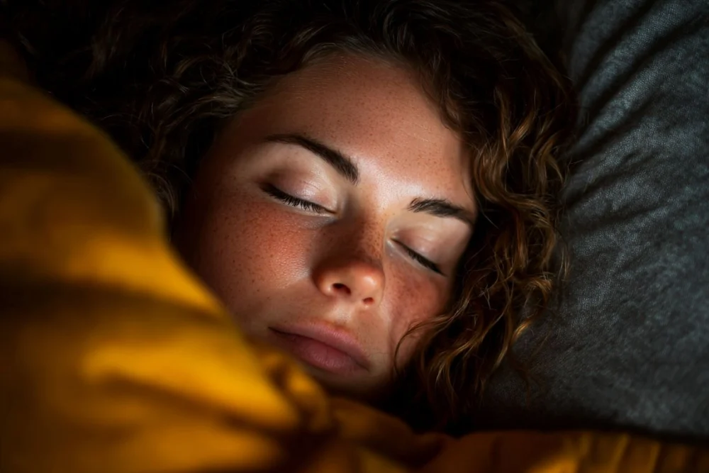
A groundbreaking study has revealed that during the deepest stages of non-REM sleep, the human brain’s activity becomes remarkably independent of the rhythmic patterns of breathing. This discovery, spearheaded by a team from Hackensack Meridian Health and its Center for Discovery and Innovation (CDI), offers profound new insights into the intricate mechanics of sleep, anesthesia, and potentially, the pathophysiology of neurological conditions like Parkinson’s disease. Published in The Journal of Neuroscience in January, the research challenges previous assumptions about the continuous coupling of vital physiological rhythms with brain activity across all sleep states, marking a significant step forward in understanding the elusive secrets held within the sleeping brain.
The Deep Dive: Unraveling Brain-Breathing Coupling
For decades, scientists have known that the brain and various bodily functions maintain a complex interplay, a phenomenon often referred to as "coupling." During wakefulness, our brains are acutely aware of and responsive to internal and external stimuli, including the rhythm of our own breathing. This coupling ensures that physiological processes are harmonized with cognitive states. Even during lighter stages of sleep, a degree of this brain-body synchronization persists. However, the latest findings suggest a fundamental shift occurs when the brain descends into the profound depths of non-REM (NREM) sleep, characterized by slow delta waves. Here, the brain appears to "disconnect" from the constant input of respiratory signals, operating on its own internal cadence.
The study, led by Bon-Mi Gu, Ph.D., also of the Hackensack Meridian School of Medicine, focused on a critical deep-brain region known as the substantia nigra. This small yet mighty area is a central hub for dopamine production and plays an indispensable role in motor control, reward, and various cognitive functions. While its importance in wakefulness and movement disorders like Parkinson’s disease is well-established, its precise role and interaction with other bodily rhythms during different sleep stages had remained largely unexplored. The research team, which includes Kolsoum Dehdar, Ph.D., and Elliot Neuberg, recently transitioned to the CDI, underscoring a concentrated effort to advance neuroscience research.
"In this study, we provide the first detailed characterization of respiration-neural coupling across multiple states – including quiet wakefulness, non-REM sleep, REM sleep, and anesthesia – in the substantia nigra and the primary motor cortex, two regions not previously studied in this context," the authors articulated in their paper. This meticulous investigation represents a crucial expansion of our understanding of how distinct brain regions behave under varying physiological conditions, moving beyond merely observing surface-level brain activity.
Methodology: Peering into the Sleeping Brain
To arrive at their conclusions, the Hackensack Meridian Health team employed sophisticated methodologies to observe the nuanced interactions between brain activity and breathing patterns in mice. Utilizing simultaneous recordings of local field potentials (LFPs) from both the primary motor cortex (M1) and the substantia nigra pars reticulata (SNr), alongside diaphragm muscle activities, researchers could precisely measure electrical brain activity and compare it with the animals’ respiratory rhythms. The experiments encompassed various states: quiet wakefulness, non-rapid eye movement (NREM) sleep, rapid eye movement (REM) sleep, and even under ketamine/xylazine anesthesia. This comprehensive approach allowed for a direct comparison of the "coupling" strength under different levels of consciousness and physiological states.
The choice of animal model, mice, provides a critical window into fundamental neural mechanisms that are often conserved across mammalian species. While direct extrapolation to human physiology always requires caution, such studies lay the foundational groundwork for future human-centric research. The ability to precisely control and monitor sleep stages, administer anesthetics, and perform invasive neural recordings in animal models offers a level of detail not always feasible in human studies.
Decoupling in Delta: The Findings Explained
The most striking discovery emerged during the deepest phases of NREM sleep. Here, the scientists observed a significant attenuation, or weakening, of the coupling between respiration and neural activity in both the substantia nigra and the primary motor cortex. This decoupling was particularly pronounced during periods of "slow delta" activity – the hallmark brain waves associated with deep, restorative sleep. Slow delta waves, typically ranging from 0.5 to 2 Hz, are indicative of widespread synchronous neural activity and are crucial for memory consolidation and physical restoration.
"The strength of respiration-neural coupling varied across multiple states, including NREM sleep, REM sleep, quiet wakefulness, and anesthesia, and was directly related to the delta power, a hallmark of NREM sleep," the authors noted. This direct correlation suggests that the very mechanisms driving deep sleep also contribute to this neural independence from respiratory input. During REM sleep and quiet wakefulness, the coupling remained stronger, indicating a more integrated brain-body system in those states.
Interestingly, the study also provided a comparative analysis with anesthesia. Under ketamine/xylazine anesthesia, the coupling was markedly enhanced in the substantia nigra, a stark contrast to natural deep sleep. This differential response highlights a fundamental distinction between pharmacologically induced unconsciousness and the natural, restorative state of deep sleep. While both involve a loss of consciousness, the underlying neural mechanisms appear to diverge significantly, particularly in how the brain interacts with vital peripheral rhythms. This finding alone has substantial implications for anesthesiology.
The Substantia Nigra: A Key Player in Sleep and Movement
The decision to focus on the substantia nigra (SNr) was a strategic one, yielding crucial insights. This region is a part of the basal ganglia, a group of subcortical nuclei vital for motor control, learning, and executive functions. The SNr, specifically, is known for its dopaminergic neurons, which are critical for smooth, coordinated movement. Its degeneration is a hallmark of Parkinson’s disease.
The study’s findings suggest that the substantia nigra, a region previously understood primarily in the context of movement and dopamine regulation, undergoes a unique functional shift during deep sleep. Its ability to "decouple" from respiratory rhythms in deep sleep, but to become more coupled under anesthesia, underscores its dynamic nature. This dynamic modulation of respiration-neural couplings within corticobasal ganglia circuits points to its potential coordinating role in body-brain interactions across different states of consciousness.
Implications for Anesthesia and Neurological Disorders
The ramifications of this research are multi-faceted, potentially reshaping our understanding of clinical practices and therapeutic strategies.
Advancing Anesthesia:
One of the most immediate practical implications lies in anesthesiology. General anesthesia aims to induce a controlled, reversible state of unconsciousness, pain relief, and muscle relaxation. However, the precise monitoring of a patient’s depth of anesthesia remains a challenge. Current methods often rely on broad indicators like heart rate, blood pressure, and sometimes EEG (electroencephalogram) readings. The discovery that natural deep sleep and anesthetic-induced unconsciousness manifest different brain-respiration coupling patterns, particularly in the substantia nigra, could pave the way for more refined monitoring techniques.
If specific patterns of decoupling or enhanced coupling in deep brain regions can reliably differentiate between natural sleep and ideal anesthetic depth, clinicians could develop more precise and personalized anesthetic protocols. This could lead to safer surgeries, minimize the risk of intraoperative awareness or excessive sedation, and improve post-operative recovery by better mimicking natural restorative processes. Understanding how anesthetics disrupt or enhance this coupling could also guide the development of new pharmacological agents that induce unconsciousness more physiologically.
A New Avenue for Parkinson’s Disease:
Perhaps even more profound are the potential implications for neurological disorders, particularly Parkinson’s disease (PD). PD is a progressive neurodegenerative disorder primarily affecting motor systems, characterized by tremors, rigidity, bradykinesia (slow movement), and postural instability. A hallmark of the disease is the loss of dopamine-producing neurons in the substantia nigra. Beyond motor symptoms, sleep disturbances are incredibly common and debilitating in PD patients, often preceding motor symptoms by years. These can include insomnia, REM sleep behavior disorder (acting out dreams), restless legs syndrome, and excessive daytime sleepiness.
The current study’s focus on the substantia nigra and its respiration-neural coupling during sleep offers a novel perspective on these sleep disruptions. If the substantia nigra’s normal decoupling mechanism during deep sleep is impaired in PD patients, it could explain some of their persistent sleep problems. A failure to "close the curtains" and allow the brain to operate independently of respiratory input might lead to fragmented, non-restorative sleep. Furthermore, if the basal ganglia’s communication with the motor cortex is altered during deep sleep in PD, as suggested by the observed SNr-M1 synchronization associated with slow delta, it could shed light on both sleep and motor control issues.
Elucidating the precise mechanisms underlying respiration-neural coupling within basal ganglia circuits could unlock new therapeutic targets for PD. For instance, interventions aimed at restoring proper decoupling in the substantia nigra during deep sleep might alleviate sleep disturbances, potentially improving overall quality of life and even slowing disease progression. This research could lead to pharmacological or even deep brain stimulation strategies tailored to modulate these specific neural rhythms during sleep.
The Broader Landscape of Sleep Science
This study contributes significantly to the broader field of sleep science, which continues to unravel the mysteries of why we sleep and what functions sleep serves. While we know deep sleep is vital for memory consolidation, waste product clearance from the brain, and cellular repair, the precise neural orchestrations underlying these processes are still being mapped. The finding that the brain actively disengages from a fundamental physiological rhythm like breathing during its deepest restorative state underscores the brain’s priority during NREM sleep: internal maintenance and processing, free from external (or even internal peripheral) distractions.
This research aligns with a growing body of evidence suggesting that sleep is not merely a passive state of rest but an active, complex process involving intricate neural reorganizations. The dynamic nature of respiration-neural coupling across different sleep stages and states of consciousness highlights the brain’s remarkable adaptability and the precise regulation required for optimal function.
Future Directions and Unanswered Questions
As with all groundbreaking research, this study opens more doors for inquiry than it closes. Several key questions and future research directions emerge:
- Human Studies: The immediate next step would be to investigate if similar respiration-neural decoupling occurs in the human substantia nigra during deep sleep. While direct invasive recordings are rare, advanced fMRI or other neuroimaging techniques could potentially reveal analogous patterns.
- Specific Neural Pathways: What are the precise neural circuits and neurotransmitter systems responsible for mediating this decoupling? Is dopamine directly involved, given its production in the substantia nigra?
- Causality vs. Correlation: Is the decoupling a cause or a consequence of deep sleep? Does actively promoting decoupling enhance the restorative qualities of sleep, or is it merely an indicator of deep sleep?
- Biomarkers: Could the strength of respiration-neural coupling serve as a biomarker for sleep quality or the presence and progression of neurological disorders?
- Therapeutic Interventions: How can this knowledge be translated into new treatments? Could non-invasive brain stimulation techniques be developed to modulate this coupling in patients with sleep disorders or Parkinson’s disease?
- Other Deep Brain Regions: Do other basal ganglia nuclei or deep brain structures exhibit similar dynamic coupling patterns during sleep and anesthesia?
Conclusion
The work by the Hackensack Meridian Health team represents a pivotal advancement in neuroscience. By meticulously observing the dynamic relationship between breathing and brain activity in the substantia nigra, they have unveiled a sophisticated mechanism of disengagement that characterizes deep sleep. This newfound understanding not only demystifies a facet of our most profound resting state but also offers tangible pathways for improving medical interventions. From refining the art of anesthesia to developing innovative therapies for debilitating conditions like Parkinson’s disease, the implications are vast. As scientists continue to delve into these intricate brain-body interactions, the promise of unlocking a healthier future through a deeper comprehension of sleep grows ever stronger. The brain, it seems, has its own unique way of tuning out the world, and even the body, to truly rest and repair.