In a significant leap forward for neuroscience and medical technology, researchers at Penn State University have developed a groundbreaking approach to 3D printing soft, stretchable bioelectrodes that are custom-tailored to the intricate and unique topography of an individual’s brain. This innovation addresses a long-standing challenge in neural interfacing: the inherent mechanical mismatch between rigid, "one-size-fits-all" implants and the delicate, highly variable structure of human brain tissue. These novel hydrogel-based sensors promise unprecedented connectivity, superior signal quality, and reduced risk of tissue damage, heralding a new era for monitoring and treating neurodegenerative diseases.
The Intricate Landscape of the Human Brain: A Challenge for Conventional Implants
The human brain, a marvel of biological engineering, is characterized by its complex surface of ridges (gyri) and grooves (sulci), a process known as gyrification. This folding dramatically increases the cortical surface area, allowing for a vast network of neurons to communicate efficiently within the confines of the skull. While the major cortical folds are generally consistent across individuals, the precise layout of these gyri and sulci varies substantially from person to person, influenced by factors such as height, weight, age, and sex. This individual variability presents a critical hurdle for traditional neural implants.
For decades, neural interfaces, which rely on tiny bioelectrodes to track biophysical signals, have predominantly utilized stiff materials with standardized designs. These conventional implants, often fabricated using lithographic techniques geared for mass production, struggle to conform intimately to the brain’s unique and dynamic three-dimensional structure. This mechanical incongruity can lead to several adverse outcomes: poor electrode-tissue contact, resulting in signal loss and unreliable data; potential damage to sensitive brain tissue due to rigidity and friction; disruption of crucial fluid transport pathways around the brain; and the elicitation of inflammatory foreign body responses, which can encapsulate the implant and further degrade its performance over time.
"Each person has a different brain structure, depending on their height, weight, age, sex and more," explained Tao Zhou, Wormley Family Early Career Professor and assistant professor of engineering science and mechanics at Penn State, and the corresponding author on the study. "Despite this, we try to fit neural interfaces onto brains like they have identical structures. This motivated us to create electrodes that are tailored for each individual, based on the structure of their brain." This fundamental understanding underscored the necessity for a paradigm shift in neural interface design.
A New Blueprint for Brain Interfaces: The Penn State Innovation
The Penn State team’s breakthrough, detailed in a paper published in Advanced Materials, lies in an integrated platform that synergizes advanced imaging, computational modeling, and sophisticated 3D printing techniques. Their approach enables the fabrication of soft, stretchable bioelectrodes that can precisely match the patient-specific geometry of the cerebral cortex.
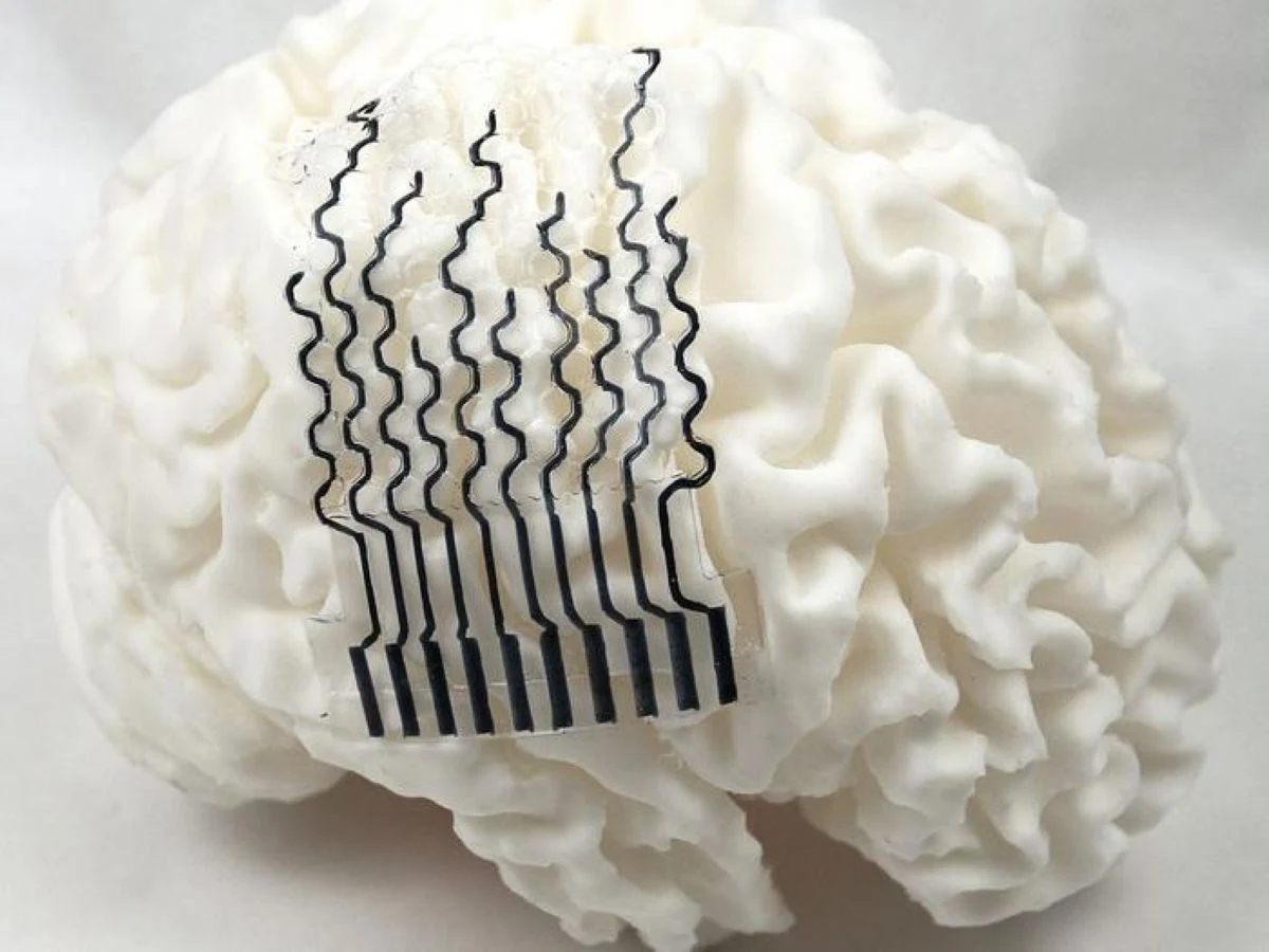
At the heart of this innovation are two key elements: the material composition and the structural design. The electrodes are primarily constructed from hydrogel, a water-rich material chosen for its exceptional softness and biocompatibility. Hydrogels possess mechanical properties that closely mimic those of brain tissue, minimizing the mechanical mismatch that plagues rigid implants. This "tissue-like" quality allows the electrodes to "morph" and stretch with the natural movements of the brain, ensuring stable and intimate contact without causing irritation or damage.
Complementing the hydrogel material is a novel honeycomb-inspired architecture. This bio-inspired design, known for its optimal strength-to-weight ratio in nature, provides the electrodes with remarkable flexibility and structural integrity. The honeycomb structure significantly reduces the overall stiffness of the electrodes while maintaining their mechanical strength, allowing them to conform deeply into the brain’s sulci without buckling or breaking. "The honeycomb structure helps us significantly reduce the stiffness of the electrodes, without sacrificing their mechanical strength," Zhou elaborated. "What’s more, the structure helps us reduce the overall material used during fabrication, reducing production time, cost and environmental impact."
From MRI to Custom Electrode: The Fabrication Pipeline
The journey from a patient’s unique brain to a custom-fit bioelectrode begins with high-resolution magnetic resonance imaging (MRI) scans. These scans provide detailed anatomical data of an individual’s brain, capturing the precise layout of their gyri and sulci. This raw data is then fed into a computational pipeline involving finite element analysis (FEA). FEA is a powerful numerical method used to simulate how a given object, under specific conditions, will react to forces and stresses. In this context, it allows researchers to create a detailed virtual model of the patient’s neural structure and predict how an electrode would interact with it, enabling optimization of the electrode’s shape and material properties for an ideal fit.
Once the optimal design is computationally derived, computer-aided design (CAD) software is used to tailor a bioelectrode specifically morphed to fit the intricate ridges and grooves of the cerebral cortex. This digital blueprint then moves to the fabrication stage, utilizing direct ink writing (DIW) 3D printing. DIW is an additive manufacturing technique where a viscous "ink" (in this case, the hydrogel material) is extruded through a nozzle to build up a 3D structure layer by layer. This method is particularly well-suited for creating complex, customized geometries with soft materials, offering significant advantages over traditional cleanroom fabrication methods.
"Traditional fabrication approaches require specialized facilities like clean rooms, making them incredibly expensive to customize," Zhou highlighted. "3D printing allows the team to personalize and manufacture electrodes much faster, for a fraction of the price." This cost-effectiveness and speed of production are crucial for making patient-specific neural interfaces a practical reality. For their study, the team 3D printed models of 21 different participant brains and physically measured how accurately their custom-designed electrodes could fit the brain surface, demonstrating superior conformability compared to conventional designs.
Unprecedented Performance and Biocompatibility
The rigorous testing of these novel bioelectrodes has yielded exceptionally promising results. Compared to traditional rigid implants, the hydrogel-based electrodes exhibited nearly perfect connectivity to the electrical signals present in the brain. The intimate and stable contact facilitated by their malleability and custom fit translates directly into higher-quality, more reliable monitoring of neural activity. This enhanced signal quality is paramount for accurate diagnosis, precise therapy delivery, and robust control of neuroprosthetic devices.
Crucially, the softness and conformability of these electrodes mean they can be applied to delicate brain tissue without causing damage, a stark contrast to the potential for injury posed by stiff conventional materials. Furthermore, the researchers confirmed that their bioelectrodes do not impede fluid transport around the brain, a vital aspect of brain function that many traditional electrodes inadvertently disrupt. Maintaining the natural physiological environment around the implant is essential for long-term integration and minimal adverse reactions.
To assess long-term biocompatibility and performance, the team conducted in vivo studies, implanting the electrodes onto the brains of rat models over a period of 28 days. The results were highly encouraging: the rats exhibited no discernible immune response to the printed electrodes, a critical benchmark in the development of any implantable biodevice. Moreover, the electrodes maintained their performance throughout the study, offering sensitive and accurate readings of the electrical and physiological signals in the rat brains without degradation. This sustained functionality and lack of inflammatory reaction underscore the potential for these personalized implants to offer durable and safe solutions for chronic applications.
Implications for Neurodegenerative Diseases and Beyond
The development of patient-specific, soft bioelectrodes represents a transformative step for a wide range of neurological applications, particularly in the realm of neurodegenerative diseases. Conditions such as Parkinson’s disease, epilepsy, Alzheimer’s disease, and multiple sclerosis could profoundly benefit from more precise monitoring and targeted therapeutic interventions.
For patients suffering from epilepsy, for instance, accurate mapping of seizure foci is critical for surgical planning. These custom-fit electrodes could provide higher-resolution, more stable recordings of epileptic activity, leading to more effective surgical resections and improved seizure control. In Parkinson’s disease, deep brain stimulation (DBS) is a well-established therapy. Personalized electrodes could potentially optimize the placement and stimulation parameters, leading to better symptom management and fewer side effects by precisely targeting specific brain regions. Similarly, for conditions like Alzheimer’s, which involve subtle and diffuse changes in brain activity, enhanced monitoring could aid in early diagnosis and tracking disease progression.
Beyond disease management, this technology holds immense promise for the advancement of neuroprosthetics and brain-computer interfaces (BCIs). BCIs aim to restore lost motor function, communication, or sensory perception by directly connecting the brain to external devices. The ability to create more stable, high-fidelity connections between the brain and prosthetics could lead to more intuitive, responsive, and reliable control of artificial limbs, wheelchairs, or communication devices, significantly improving the quality of life for individuals with severe disabilities.
Zhou envisions a future where this printing method serves as a robust framework for the commercial-scale production of customized bioelectrodes. "We are looking to further improve this technology to optimize the electrodes to monitor for specific diseases," Zhou stated. "In the future, we would really like to work with patients to see how this approach could support brain monitoring and disease treatment in clinical settings." This progression towards clinical trials and eventual widespread adoption underscores the significant potential impact of this research. The personalized approach aligns perfectly with the growing trend in medicine towards individualized treatments, where therapies are tailored to the unique biological and physiological characteristics of each patient.
The Future of Personalized Neurology
The Penn State research team’s pioneering work marks a critical juncture in the evolution of neural interface technology. By addressing the fundamental challenge of anatomical variability and mechanical mismatch, they have paved the way for a new generation of implants that are not only more effective but also safer and more comfortable for patients. The ability to rapidly and cost-effectively 3D print custom hydrogel electrodes opens up unprecedented possibilities for personalized neurology, moving beyond generalized solutions to therapies precisely tuned to the individual brain.
This groundbreaking research was supported by the U.S. National Science Foundation and the National Institutes of Health, reflecting the recognized importance of advancing neural interface capabilities. The collaborative effort involved a multidisciplinary team from Penn State, including Nanyin Zhang, professor of biomedical engineering; Sulin Zhang, professor of engineering science and mechanics; and a cohort of dedicated doctoral candidates and researchers across engineering science, mechanics, and biomedical engineering. Their collective expertise has culminated in an innovation that stands to redefine how we interact with, understand, and treat the most complex organ in the human body. As this technology matures and moves closer to clinical application, it promises to unlock new avenues for improving neurological health and enhancing human capabilities.